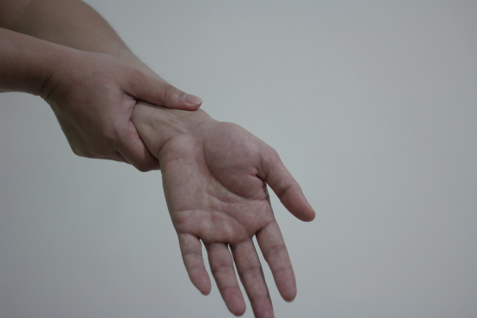
The wrist is made up of 8 small carpal bones arranged in two rows. The scaphoid bone is an obliquely oriented bone that can be found on the radial (thumb) side of the wrist. It is the most commonly fractured carpal bone in the hand accounting for 60-70% of all hand fractures. The majority of the bone (up to 80%) is covered by articular cartilage, which leaves very little room for blood supply to the bone and ligamentous attachments. The most common way this bone is injured is during a fall on an outstretched hand, also known as a FOOSH-type injury. Males aged 20-24 seem to fracture their scaphoid bones at a higher rate, along with individuals who play high impact or extreme sports such as motocross, basketball, and football. It is estimated that 1 in every 100 football athletes in college football will at one point suffer a scaphoid fracture2.
Common Symptoms
Despite the common nature of these fractures, they tend to be quite hard to diagnose. Patients will often present with pain and swelling in an area called the “snuffbox” on the thumb-side of the wrist. They often will have a loss of range of motion, and pain with both movement of the wrist, and putting weight through the wrist (getting up from the bed/chair etc.). Up to 20% are initially not found on conventional x-ray imaging. Early identification can be very important given the abnormal blood supply to this bone. Depending on the location of the fracture, there may indeed be a risk of non-union, which would lead to the need for a surgical fixation via a screw. Common practice, should a scaphoid fracture be suspected but the initial x-ray is negative, would be to cast the hand/wrist for 10-14 days and repeat the imaging. Special x-ray views are also needed to identify the entire bone. Both MRI and/or CT scans can be used for further viewing of the bone for surgical planning.
Traditionally, fractures at the distal pole of the bone, along with fractures that are “non-displaced” are treated with immobilization via a thumb spica splint/cast. Early identification and treatment are imperative to ensure proper healing and avoiding long-term dysfunction. Fractures that are treated within 4 weeks of the injury have a higher union rate than fractures treated >4 weeks from the date of injury. Healing times vary, and typically, the more proximal the fracture, the length required healing time. Typical immobilization times are ~ 8-12 weeks. However, in some cases, the bone may take upwards to 6 months to unite. Often times, fractures of the distal pole will unite in 6 weeks, versus fractures of the proximal pole taking 6 months as noted above. Prolonged immobilization comes with its own risks, including contracture of the muscles, osteopenia, and disuse of the muscles creating significant amounts of atrophy and weakness. As noted above, any fracture that is displaced, or demonstrates non-union on imaging such as x-ray, MRI or CT, may require surgical fixation via a placement of a screw.
Stay Mobile,
Travis Gaudet MScPT., BScKin., F.C.A.M.P.T., Dip.Manip.PT., cGIMS
References:
1. https://www.hand.theclinics.com/article/S0749-0712(19)30017-4/abstract
2. Winston, Mark J, and Andrew J Weiland. “Scaphoid Fractures in the Athlete.” Current Reviews in Musculoskeletal Medicine, Springer US, www.ncbi.nlm.nih.gov/pmc/articles/PMC5344853/.


